Apoptosis, not so quiet after all

Posted by Kasra

Apoptosis has been conventionally regarded as a quiet and non-inflammatory event, compared to necrosis which results in release of alarmins and other danger signals inducing inflammation and immune cell recruitment. However, a recent report by Cullen et al. published in Molecular Cell suggests otherwise. They suggest that at least in one form of apoptosis, pro-inflammatory mediators are released by the apoptotic cells and can act as ‘find-me’ signals to the phagocytes to find and clear them.

Fas receptor or CD95 is among the famous apoptosis receptors. It is a member of the TNF receptor family and it induces apoptosis through Caspase-8 activation. Interestingly, Cullen et al. show that different cell types produce pro-inflammatory chemikones such as MCP-1, CXCL1 and MIP-2 when they go through apoptosis via Fas-pathway. They show that this chemokine release is NF-kappa-B mediated and independent of Caspase-8 activation. It is possible be that somewhere during the evolution, the apoptotic pathway cross-linked with the pro-inflammatory signaling pathway and found benefit in it. Accordingly, Cullen et al. show that the Fas-induced pro-inflammatory cytokine/chemokine production still occurs  even if the apoptotic pathway is inhibited showing that these pathways are separate.

Next the authors show that the supernatant from the apoptotic cells can induce migration of macrophages and neutrophils. They also pinpoint the responsible chemokine by depleting them one-by-one. They show that MCP-1 induces macrophage migration and IL-8 recruits neutrophils.

At this point it cannot be said really how inflammatory these apoptotic cells would be in vivo. There are parts of the body where apoptosis occurs constantly,  so this could potentially lead to an unwanted constant inflammation in those areas. Therefore, either different cells would have different levels of apoptotic-proinflammatory chemokine release, or local mechanisms would compensate and counteract the inflammation. More studies will help us understand how apoptotic cells can send their ‘find-me’ signals without causing too much turbulence in their tissue.

Schematic diagram of model proposed by Cullen et al. From

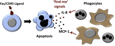
From Cullen et al. Molecular cell, 49 (6), 1034-48

Cullen SP, Henry CM, Kearney CJ, Logue SE, Feoktistova M, Tynan GA, Lavelle EC, Leverkus M, & Martin SJ (2013). Fas/CD95-induced chemokines can serve as “find-me” signals for apoptotic cells. Molecular cell, 49 (6), 1034-48 PMID: 23434371

ResearchBlogging.org

We know Nirtic Oxide is produced when SHP-1 is absent, but why??

Posted by: Issa Abu-Dayyeh

The exact role played by the protein tyrosine phosphatase (SHP-1) in the negative regulation of macrophage functions has been an active area of research for many years. In fact, SHP-1 deficient mice are hyper-inflammatory. They lose their hair “for God’s sake” due to exaggerated inflammatory responses in the skin area! (hence their name motheaten). But what does this tell us? It tells us a lot of pathways are simply “on fire”. To dissect every single pathway controlled by this PTP is a humongous job, and the best approach in my opinion is to try to focus, and dissect a pathway at a time and a function at a time. So, what did we attempt to do in our most recent publication (Blanchette, J. et al.) in Immunology (2008)?

The paper explores the signaling pathways that seem to be major contributors to NO production in SHP-1 deficient macrophages. NO production is driven by a gene known as iNOS whose expression is driven by several transcription factors, most importantly: Nf-kB, STAT, and AP-1. One of those transcription factors “AP-1” is activated by a MAP kinase called JNK.

This work utilizes inhibitors of many of these members to see which of them will be able to suppress that excess NO production observed in SHP-1 deficient macrophages in an effort to understand how SHP-1 causes this increased NO production.

To save you the dull experimental details…Results showed that the exaggerated NO production in SHP-1-/- macrophages seems to be due to an increased JNK/AP-1 and not NF-kB activity.

And so what? some people might ask!

Well…I agree a finding like this might not find a cure to leishmaniasis. Nevertheless, bearing in mind that NF-kB translocation is increased in the absence of SHP-1, this paper then suggests something rather important. This increased NF-kB activity is not contributing to iNOS transcription. What is it doing then? and how can iNOS be differentially regulated? These are questions that await answers. (If somebody has answers, I will be glad to hear from them).

This work simply broadens our knowledge about where SHP-1 exerts its effects, and by knowing how, we can probably try to eventually revert some of those actions during the course of a Leishmania infection and help find an effective drug against leishmaniasis that is not as toxic as the ones available nowadays…

A block added to the wall. that is how I see it.

If you are interested in viewing the paper, please visit it here

Enjoy,

Issa